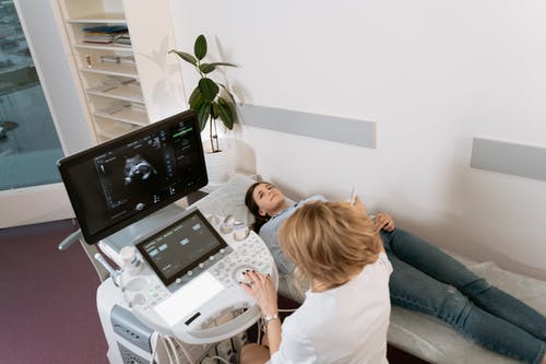
Ultrasound technology has come a long way from its humble beginnings as a tool for monitoring pregnancy. Today, it is one of the most versatile and commonly used imaging techniques in modern medicine. Ultrasound provides real-time, non-invasive imaging, and it has found applications in various specialties, from obstetrics and cardiology to musculoskeletal and emergency medicine.
In this blog post, we’ll break down the different types of ultrasound imaging techniques—such as 2D, 3D, and Doppler ultrasound—and explore how each is used to diagnose a range of medical conditions.
1. 2D Ultrasound (Two-Dimensional Ultrasound)
What Is It? 2D ultrasound, the most common type of ultrasound imaging, produces a flat, two-dimensional image of the internal organs and tissues. This technique uses high-frequency sound waves that bounce off the body’s tissues and return to the ultrasound machine, creating an image of what is beneath the surface.
Applications of 2D Ultrasound:
- Obstetrics and Gynecology: 2D ultrasound is widely used in prenatal care to monitor fetal development, measure the baby’s growth, and check for potential complications like ectopic pregnancies or placental issues. It’s typically used during routine check-ups to assess the fetus and the health of the mother.
- Abdominal Imaging: 2D ultrasound is also commonly used to examine the organs in the abdominal area, such as the liver, kidneys, gallbladder, and pancreas. This helps identify conditions like liver disease, gallstones, and kidney abnormalities.
- Cardiology: In cardiology, 2D echocardiography is used to view the heart’s structure, its chambers, and the valves, helping diagnose heart disease, valve issues, and other cardiovascular conditions.
Advantages of 2D Ultrasound:
- Non-invasive and safe: Unlike other imaging techniques like X-rays or CT scans, 2D ultrasound doesn’t use radiation, making it safe for both patients and healthcare providers.
- Real-time imaging: It provides live, moving images, which is especially useful for monitoring the heart, blood vessels, and organs in motion.
2. 3D Ultrasound (Three-Dimensional Ultrasound)
What Is It? 3D ultrasound takes the basic principles of 2D ultrasound and adds an extra layer of depth, creating a three-dimensional image. The technology captures multiple 2D images from different angles and then combines them to create a 3D image, offering more detailed views of structures and organs.
Applications of 3D Ultrasound:
- Obstetrics: One of the most popular applications of 3D ultrasound is in prenatal care. This technique provides a clearer and more detailed image of the fetus, allowing parents to see their baby’s facial features or even determine the baby’s gender. It’s often used in elective imaging, providing a more engaging experience for parents-to-be.
- Gynecology: In gynecology, 3D ultrasound can help visualize the uterus, ovaries, and other reproductive organs in greater detail. It’s used to assess conditions like uterine fibroids, ovarian cysts, and structural abnormalities.
- Cardiology: 3D ultrasound (also known as 3D echocardiography) is increasingly used in cardiology to provide more precise images of the heart’s anatomy, which is beneficial for assessing congenital heart defects, valve function, and blood flow.
Advantages of 3D Ultrasound:
- Detailed and clear images: 3D ultrasound provides a high level of detail, offering more accurate and comprehensive views compared to 2D ultrasound.
- Better visualization of complex structures: The added depth in 3D imaging is particularly helpful for viewing complex structures, such as organs or the heart, in a way that can help doctors make more accurate diagnoses.
3. Doppler Ultrasound
What Is It? Doppler ultrasound is a specialized technique that measures the flow of blood within blood vessels. It uses sound waves to assess the movement of blood cells and can show the speed and direction of blood flow. This technique is particularly useful for assessing the circulatory system.
Applications of Doppler Ultrasound:
- Vascular Studies: Doppler ultrasound is commonly used to examine blood flow through arteries and veins. It’s often used to detect conditions like blood clots, deep vein thrombosis (DVT), and narrowing or blockages in blood vessels (such as in carotid artery disease or peripheral artery disease).
- Cardiology: Doppler echocardiography is used to evaluate heart function by assessing blood flow through the heart’s chambers and valves. It helps detect issues like heart valve defects, heart failure, and congenital heart conditions.
- Obstetrics: Doppler ultrasound can be used in pregnancy to monitor the blood flow between the placenta and fetus. It helps evaluate the well-being of the baby and detects potential issues like growth restriction or placental insufficiency.
Advantages of Doppler Ultrasound:
- Real-time blood flow data: Doppler ultrasound provides real-time data on blood flow, making it invaluable for diagnosing vascular and heart-related issues.
- Non-invasive and quick: Like other ultrasound techniques, Doppler ultrasound is non-invasive and can provide results quickly, without the need for more invasive procedures like angiography.
4. 4D Ultrasound (Four-Dimensional Ultrasound)
What Is It? 4D ultrasound is essentially an extension of 3D ultrasound. While 3D ultrasound provides still images, 4D ultrasound captures moving images, offering real-time video of the fetus or other structures. It creates a more dynamic view, often used for prenatal imaging.
Applications of 4D Ultrasound:
- Obstetrics: 4D ultrasound is often used in prenatal care to offer a more engaging and realistic view of the fetus. It can show live movement of the baby, including facial expressions, hand movements, and even yawning or hiccuping, which is exciting for expectant parents.
- Therapeutic and Diagnostic Use: 4D ultrasound can be used for advanced diagnostics, especially in the early stages of pregnancy, to check for abnormalities in fetal movement or physical development.
Advantages of 4D Ultrasound:
- More engaging experience: Expectant parents appreciate the ability to see their baby’s movements in real time. It also provides a more in-depth view of the fetus’s development.
- Improved visualization of fetal abnormalities: 4D ultrasound can sometimes offer a clearer picture of developmental issues that might be harder to identify with 2D or 3D imaging alone.
5. Contrast-Enhanced Ultrasound
What Is It? Contrast-enhanced ultrasound uses special contrast agents (tiny bubbles filled with gas) that are injected into the bloodstream. These contrast agents improve the ultrasound image, allowing healthcare professionals to better visualize blood vessels, organs, and tumors.
Applications of Contrast-Enhanced Ultrasound:
- Liver Disease: It can help doctors visualize liver tumors, cysts, and abnormalities more clearly, often used in patients with liver cirrhosis or hepatitis.
- Cancer Imaging: Contrast-enhanced ultrasound is also used to evaluate the vascularity of tumors, helping doctors assess whether a mass is benign or malignant.
- Cardiovascular Imaging: It can improve visualization of the heart and blood vessels, particularly in patients with heart disease.
Advantages of Contrast-Enhanced Ultrasound:
- Improved imaging in difficult-to-see areas: The use of contrast agents enhances ultrasound images, making it easier to detect abnormalities that may not be visible with standard ultrasound techniques.
- Faster than other contrast techniques: Unlike CT or MRI, contrast-enhanced ultrasound is faster, often making it a preferred option in emergency or outpatient settings.
Conclusion
Ultrasound technology has come a long way, offering a variety of techniques to meet the needs of modern healthcare. From the simplicity of 2D ultrasound to the advanced capabilities of 3D and Doppler imaging, each ultrasound technique has specific applications that enhance diagnostic accuracy and patient care. Whether you’re monitoring fetal health, diagnosing heart disease, or evaluating blood flow, ultrasound is a versatile and non-invasive tool that continues to shape the future of medicine.
At Ultrasound Trainings, we offer comprehensive ultrasound training programs that cover these techniques and more, ensuring that you gain the skills needed to excel in the field of medical imaging. If you’re ready to take the next step in your ultrasound career, contact us today to learn more about our training programs!


