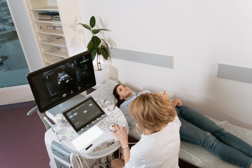
In recent years, point of care ultrasound (POCUS) has revolutionized the field of obstetrics and gynecology (OB/GYN) by providing real-time, bedside imaging to assist with immediate decision-making. POCUS allows OB/GYN practitioners to quickly assess, diagnose, and manage a wide range of conditions, from early pregnancy complications to gynecologic abnormalities, without the need for immediate referral to radiology. This makes it an invaluable tool, not only in emergency settings but also in routine clinical practice.
For OB/GYN practitioners looking to maximize the use of POCUS in their practice, there are essential tips and best practices to ensure its effectiveness, safety, and diagnostic accuracy. In this detailed blog post, we will dive deep into the fundamentals of OB/GYN POCUS, key tips to optimize its use, and how it can enhance patient care.
1. Understanding the Basics of OB/GYN POCUS
Point of care ultrasound (POCUS) is a portable and non-invasive imaging tool used by OB/GYN practitioners to perform ultrasound exams at the patient’s bedside. Unlike traditional diagnostic ultrasound, which is performed by sonographers and interpreted by radiologists, POCUS is typically performed by the clinician during a patient visit, allowing for immediate results and decisions.
In OB/GYN practice, POCUS can be used for:
- Early Pregnancy Assessment: Confirming intrauterine pregnancy, detecting ectopic pregnancies, assessing gestational age, and monitoring fetal viability.
- Pelvic Ultrasound: Evaluating uterine and ovarian pathology such as fibroids, ovarian cysts, or endometriosis.
- Guiding Procedures: Assisting with the placement of an IUD, performing amniocentesis, or guiding the aspiration of ovarian cysts.
- Emergency Obstetric and Gynecologic Situations: Evaluating complications such as ruptured ectopic pregnancies, vaginal bleeding, or assessing the fetal heart rate in labor.
2. POCUS Tips for Early Pregnancy Evaluation
Early pregnancy is one of the most common uses of POCUS in OB/GYN. Here are some essential tips to help you get accurate and reliable results:
2.1 Assessing for Intrauterine Pregnancy (IUP)
When performing POCUS to evaluate early pregnancy, ensure that you can distinguish between an intrauterine pregnancy (IUP) and a possible ectopic pregnancy.
- Tip: Start by confirming the presence of a gestational sac within the uterus. Use a high-frequency transvaginal probe for early gestational sac visualization, usually around 4-5 weeks gestation.
- Tip: Look for the yolk sac within the gestational sac, as this is a more specific sign of an IUP around 5 weeks. By 6 weeks, the fetal pole should be visible with a heartbeat.
2.2 Evaluating Ectopic Pregnancy
Ectopic pregnancy can be a life threatening condition, making it critical to diagnose early.
- Tip: Always evaluate for free fluid in the pelvis, which may indicate rupture or impending rupture of an ectopic pregnancy. Even a small amount of free fluid can raise suspicion.
- Tip: If a gestational sac is seen outside of the uterus, it is highly suggestive of an ectopic pregnancy. Check for the presence of an adnexal mass, which is a common finding in ectopic pregnancies.
- Tip: When assessing suspected ectopic pregnancy, use both transabdominal and transvaginal imaging to improve diagnostic accuracy. The vaginal probe is more sensitive for early pregnancies and can provide clearer images of the uterus and adnexa.
2.3 Assessing Fetal Viability
Once the fetal pole is visible, assess the fetal heart rate (FHR) to confirm fetal viability.
- Tip: Use color Doppler or pulse Doppler to visualize and measure the fetal heart rate. A normal heart rate is between 120-160 beats per minute. If you do not see cardiac activity in a structure that appears to be a fetal pole, this may suggest a non-viable pregnancy, and further management will be needed.
3. POCUS Tips for Gynecologic Conditions
In addition to pregnancy-related uses, POCUS is also invaluable in diagnosing various gynecologic conditions, particularly when it comes to pelvic pain, abnormal bleeding, and reproductive tract abnormalities.
3.1 Evaluating Uterine Pathologies
POCUS can be used to evaluate uterine abnormalities such as fibroids, adenomyosis, and endometrial thickening.
- Tip: For fibroids, use a transabdominal or transvaginal approach, depending on the patient’s body habitus. Fibroids will appear as well-defined, hypoechoic masses within the myometrium (muscle layer of the uterus). Make sure to measure the size of the fibroids and document their location for future reference.
- Tip: If evaluating for endometrial hyperplasia or malignancy, assess the endometrial stripe for thickening. A thickened endometrium (>14mm) in postmenopausal women warrants further evaluation.
3.2 Assessing Ovarian Cysts
Ovarian cysts are common findings in reproductive-aged women and can present with pelvic pain.
- Tip: Use a high-frequency transvaginal probe for better resolution when assessing ovarian cysts. Simple cysts will appear as an anechoic (black) fluid-filled structure. Measure the cyst size and monitor for any complex features, such as septations or solid components, which may suggest malignancy or hemorrhage.
- Tip: For women with suspected torsion, look for an ovary with a twisted or abnormal appearance, along with signs of decreased blood flow on Doppler imaging.
3.3 Pelvic Free Fluid
In cases of pelvic pain or suspected rupture of an ovarian cyst, it is important to check for free fluid in the pelvis.
- Tip: Free fluid in the pelvis may suggest rupture of an ovarian cyst, ectopic pregnancy, or endometriosis. Using the Morrison’s pouch (the area between the liver and right kidney) is essential for detecting significant fluid.
4. POCUS Tips for Obstetric Emergencies
In emergency situations, such as suspected fetal distress, hemorrhage, or trauma, POCUS can provide quick insights to guide management decisions.
4.1 Assessing Fetal Heart Rate in Labor
Monitoring the fetal heart rate (FHR) in labor is critical, especially when there are concerns about fetal well-being.
- Tip: Use doppler ultrasound to measure the fetal heart rate during labor. This is particularly useful in situations where external monitoring (e.g., a cardiotocograph) is unavailable or inappropriate.
- Tip: A bradycardic fetal heart rate (<100 bpm) may be a sign of fetal distress, and immediate intervention is required.
4.2 Assessing for Placental Abruption or Previa
Placental abruption and previa are serious obstetric conditions that can result in hemorrhage.
- Tip: In cases of vaginal bleeding or abdominal pain, perform POCUS to assess for placental location and any signs of placental abruption. In placental abruption, the placenta will appear separated from the uterine wall with hypoechoic areas between the placenta and myometrium.
- Tip: For suspected placenta previa, visualize the placental position in relation to the cervical os (opening). If the placenta covers the internal os, it is diagnosed as previa, and immediate planning for delivery should be considered.
5. POCUS Tips for Guided Procedures
POCUS can be invaluable for guiding certain OB/GYN procedures, such as amniocentesis, IUD placement, or aspiration of ovarian cysts.
5.1 Guiding IUD Placement
POCUS is particularly useful when placing intrauterine devices (IUDs), as it allows the clinician to visualize the uterine cavity and confirm IUD placement.
- Tip: Use a transabdominal ultrasound to visualize the uterine cavity during IUD insertion, ensuring that the device is placed in the correct location.
- Tip: If there are any concerns about the IUD being positioned incorrectly (e.g., perforation), perform an ultrasound immediately after the procedure to verify placement.
5.2 Assisting with Amniocentesis
POCUS can be used to guide amniocentesis to ensure that the needle is inserted in the correct location and to avoid injury to the fetus or other structures.
- Tip: Use real-time visualization to confirm the needle’s trajectory and the location of the amniotic sac. Always measure the amount of amniotic fluid available to avoid complications.
6. POCUS Tips for Improving Skills and Accuracy
To ensure the accuracy and efficacy of POCUS in OB/GYN practice, clinicians must continue to build their skills and knowledge.
6.1 Maintain Regular Practice
POCUS is a skill that improves with practice, and regular use is key to increasing accuracy and confidence.
- Tip: Make POCUS a regular part of your clinical practice. The more you use it, the more skilled you will become in interpreting images and making quick decisions.
6.2 Take Continuing Education Courses
Participating in formal POCUS training programs or workshops will help you keep up with the latest techniques, guidelines, and technologies.
- Tip: Many professional societies, including the American Institute of Ultrasound in Medicine (AIUM), offer certification programs that can help you gain expertise in POCUS for OB/GYN.
Conclusion
Point of care ultrasound (POCUS) has become an indispensable tool in OB/GYN practice, enhancing diagnostic accuracy, streamlining decision-making, and improving patient care. By incorporating POCUS into your clinical workflow and following these key tips, you can enhance your ability to assess early pregnancy, diagnose gynecologic conditions, manage obstetric emergencies, and guide procedures with confidence. Continuous practice, further education, and collaboration with experienced practitioners will ensure that you can maximize the full potential of POCUS in your practice and provide the best care possible for your patients.