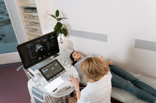
Ultrasound is one of the most versatile and accessible diagnostic tools in modern healthcare. It offers real-time imaging, portability, and safety across a wide range of clinical applications—from obstetrics and gynecology to emergency medicine, cardiology, and primary care. However, like any diagnostic technology, ultrasound has its limitations. Understanding when ultrasound is not the right tool is just as important as knowing how to use it effectively. At Ultrasound Trainings, we believe that well-informed providers deliver better care—not by relying on technology alone, but by using it appropriately and collaboratively.
Understanding What Ultrasound Can (and Can’t) Do
Ultrasound relies on the transmission and reflection of sound waves to create images. While it excels at evaluating soft tissue structures and fluid-filled spaces, it has significant limitations when sound cannot penetrate or reflect effectively. These physical and biological constraints shape how ultrasound should be used in clinical practice.
Common Limitations of Ultrasound
1. Limited Penetration Through Air or Bone
Air and bone are major barriers to ultrasound imaging. The lungs, bowel gas, and bones scatter or absorb sound waves, creating acoustic shadows that obscure deeper structures. For this reason, ultrasound is not ideal for evaluating:
- Pulmonary parenchyma (beyond pleural evaluation)
- Bowel pathology obscured by gas
- Intracranial structures (in adults with closed fontanelles)
Providers should recognize when other imaging modalities, such as CT or MRI, are more appropriate for these cases.
2. Operator Dependence
Ultrasound accuracy heavily depends on the operator’s training, experience, and technique. Poor probe placement, incorrect settings, or misinterpretation can lead to inaccurate conclusions. This variability underscores why structured training and competency evaluation—such as that offered at Ultrasound Trainings—is essential for safe practice.
3. Limited Field of View
Unlike CT or MRI, ultrasound provides a narrow, real-time “window” rather than a comprehensive image. Pathologies outside the scanned region can easily be missed. For example, ultrasound might detect a localized abscess but not reveal deeper or distant infection. Providers must integrate ultrasound findings with the overall clinical picture and follow up with additional imaging when needed.
4. Difficulty Visualizing Deep or Obese Patients
Body habitus can significantly affect image quality. Increased tissue depth attenuates sound waves, making it challenging to visualize deep organs like the pancreas, kidneys, or fetal anatomy in late gestation. Adjusting machine settings and using lower-frequency probes can help, but sometimes another imaging method will provide more diagnostic clarity.
5. Non-Specific Findings
Many ultrasound findings are non-specific. For example, a “complex ovarian cyst” could represent several different diagnoses—from a benign hemorrhagic cyst to an endometrioma or malignancy. In such cases, ultrasound serves as a starting point, guiding further evaluation with other imaging modalities or laboratory testing.
6. Not a Substitute for Comprehensive Evaluation
Ultrasound should never replace a full clinical assessment. It’s a tool to enhance—not replace—clinical reasoning. Over-reliance on imaging can lead to missed diagnoses or unnecessary reassurance. A normal scan does not always equal a normal patient.
When to Consider Other Imaging Modalities
- CT Scans: Excellent for evaluating bone, lung, trauma, and detailed cross-sectional anatomy.
- MRI: Preferred for soft tissue differentiation, brain imaging, and cases requiring no radiation exposure but more detail than ultrasound provides.
- X-ray: Ideal for skeletal imaging and quick assessments of the chest and abdomen where ultrasound is limited by air.
Each imaging modality has its place. The key is knowing which tool provides the right information for the clinical question at hand.
Why Ultrasound Training Still Matters
Even with its limitations, ultrasound remains one of the most useful bedside diagnostic tools available. When providers understand both its capabilities and its boundaries, they can use it to enhance—not compromise—patient care. At Ultrasound Trainings, our education goes beyond technical skills to include image interpretation, safety principles, and clinical decision-making.
Our goal is to create confident, competent providers who know when ultrasound adds value—and when another tool is a better choice.
Ready to deepen your ultrasound expertise? Explore our hands-on workshops and advanced training programs at Ultrasound Trainings. Learn how to use ultrasound safely, effectively, and wisely—because true mastery means knowing when not to scan.


