Uterine fibroids are non-cancerous growths that develop in or on the uterus. Also known as leiomyomas or myomas, fibroids are common in women of reproductive age and can vary in size and number. While many fibroids are asymptomatic, others may cause symptoms such as heavy menstrual bleeding, pelvic pain, and reproductive issues.
Ultrasound is one of the most effective imaging techniques for diagnosing and monitoring fibroids. It is non-invasive, safe, and provides detailed images of the uterus and fibroid growth.
What Are Uterine Fibroids?
Fibroids are benign tumors composed of smooth muscle and fibrous tissue. They can develop in different areas of the uterus, and their size can range from as small as a pea to as large as a melon. There are several types of fibroids, categorized based on their location within the uterus:
- Intramural Fibroids: These grow within the muscular wall of the uterus and are the most common type.
- Subserosal Fibroids: These develop on the outer surface of the uterus and may cause the uterus to appear larger on one side.
- Submucosal Fibroids: Located under the inner lining of the uterus, these can extend into the uterine cavity and are often associated with heavy menstrual bleeding.
- Pedunculated Fibroids: These fibroids grow on a stalk inside or outside the uterus.
While fibroids are generally non-cancerous, they can cause significant symptoms in some women, such as:
- Heavy or prolonged periods
- Pelvic pain and pressure
- Frequent urination
- Difficulty emptying the bladder
- Pain during intercourse
- Reproductive issues, including infertility or pregnancy complications
Why Is Ultrasound Used for Fibroids?
Ultrasound is the first-line imaging tool for diagnosing and evaluating uterine fibroids. It is widely available, cost-effective, and does not involve radiation exposure. Using sound waves to create images, ultrasound allows healthcare providers to assess the size, location, and number of fibroids, providing crucial information for treatment planning.
Advantages of Ultrasound for Fibroid Diagnosis:
- Non-invasive and painless
- Provides real-time imaging
- Safe for pregnant women and those planning to conceive
- Detects fibroids of various sizes and types
Types of Ultrasound Used for Fibroid Evaluation
There are two main types of ultrasound used to evaluate fibroids: transabdominal ultrasound and transvaginal ultrasound. In some cases, both types may be used to get a comprehensive view of the uterus and the fibroids.
1. Transabdominal Ultrasound
A transabdominal ultrasound involves placing a transducer on the abdomen to capture images of the uterus. The transducer emits sound waves that bounce off the structures within the pelvis, creating an image of the uterus and fibroids on a monitor.
This type of ultrasound provides a wider view of the pelvic area, making it useful for detecting large fibroids or those located on the outer surface of the uterus. However, it may not provide as detailed a view of smaller fibroids or those located deep within the uterine wall.
2. Transvaginal Ultrasound
A transvaginal ultrasound involves inserting a small, lubricated transducer into the vagina. This allows for a closer and more detailed view of the uterus and fibroids, especially those located inside the uterine cavity or within the muscular wall. Transvaginal ultrasound is often more sensitive in detecting smaller fibroids and in assessing the endometrial lining, which is important when submucosal fibroids are suspected.
What Does an Ultrasound Reveal About Fibroids?
Ultrasound imaging provides detailed information about fibroids, including:
1. Size of Fibroids
The size of fibroids can vary significantly, from as small as a seed to as large as a grapefruit. Ultrasound measurements help determine the size of each fibroid, which is important for treatment decisions. Large fibroids may require more aggressive treatment, while smaller ones may only need monitoring.
2. Location of Fibroids
Ultrasound identifies where the fibroids are located within the uterus, which helps guide treatment. For example, submucosal fibroids that protrude into the uterine cavity can cause heavy bleeding and may require removal, while intramural fibroids growing within the uterine wall may cause pain and reproductive issues.
3. Number of Fibroids
Fibroids can occur as a single growth or multiple masses. Ultrasound can count the number of fibroids, which helps determine the complexity of the condition. Multiple fibroids may be managed differently than a single large fibroid, and ultrasound provides an accurate count.
4. Characteristics of Fibroids
Ultrasound can distinguish between fibroids and other conditions that affect the uterus, such as adenomyosis or endometrial polyps. By providing details about the texture and appearance of the fibroids, ultrasound helps confirm the diagnosis and rule out other potential issues.
5. Effects on Surrounding Organs
Large fibroids can press against surrounding organs, such as the bladder or bowel, leading to symptoms like frequent urination or constipation. Ultrasound can show how the fibroids are affecting nearby organs, helping to explain certain symptoms.
When Is an Ultrasound for Fibroids Recommended?
Ultrasound is recommended in several situations related to fibroids, including:
- Unexplained heavy menstrual bleeding: If a woman experiences abnormally heavy or prolonged periods, an ultrasound can help determine if fibroids are the cause.
- Pelvic pain or pressure: Ultrasound is often ordered to evaluate the source of persistent pelvic pain or pressure.
- Reproductive issues: Women struggling with infertility or recurrent miscarriages may have fibroids that affect their reproductive system, which can be detected via ultrasound.
- Monitoring known fibroids: Women with a history of fibroids may undergo regular ultrasounds to monitor any changes in size or number.
What to Expect During an Ultrasound for Fibroids
An ultrasound for fibroids is a straightforward procedure that usually takes about 15 to 30 minutes. Here’s what to expect:
Preparation
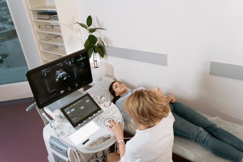
For a transabdominal ultrasound, you may be asked to have a full bladder, as this helps improve the clarity of the ultrasound images. You’ll lie on an examination table while the sonographer applies gel to your abdomen. The gel helps the transducer glide smoothly over your skin and ensures good contact for clear images.
For a transvaginal ultrasound, no special preparation is needed. You will be asked to empty your bladder before the procedure. A small transducer will be gently inserted into the vagina to capture images.
The Procedure
The sonographer will move the transducer over your abdomen (or inside the vagina for a transvaginal ultrasound) to capture images of the uterus and fibroids. You may feel some pressure, but the procedure is generally painless.
Results
The images will be analyzed by a radiologist or your doctor, who will explain the findings. If fibroids are detected, your doctor will discuss treatment options based on the size, location, and symptoms associated with the fibroids.
Treatment Options for Fibroids Based on Ultrasound Findings
The results of the ultrasound help guide treatment decisions. Some common treatment options include:
- Watchful waiting: If the fibroids are small, asymptomatic, and not affecting fertility, your doctor may recommend periodic ultrasounds to monitor for any changes.
- Medication: Hormonal treatments such as gonadotropin-releasing hormone (GnRH) agonists or birth control pills can help shrink fibroids and reduce symptoms.
- Surgical options: For larger or symptomatic fibroids, surgery may be recommended. Options include myomectomy (removal of the fibroids while preserving the uterus) or hysterectomy (removal of the uterus).
- Non-surgical treatments: Procedures like uterine fibroid embolization (UFE) can shrink fibroids by cutting off their blood supply. MRI-guided focused ultrasound is another option that uses sound waves to destroy fibroids without surgery.
Conclusion
Ultrasound is a vital tool in diagnosing and managing uterine fibroids. It provides detailed images that help doctors understand the size, location, and number of fibroids, which is crucial for determining the best treatment options. Whether you are experiencing symptoms or have known fibroids, regular ultrasounds can help monitor your condition and ensure you receive appropriate care.


