Endometriosis is a chronic and often painful condition in which tissue similar to the lining of the uterus (endometrium) grows outside the uterus, affecting the ovaries, fallopian tubes, and other pelvic organs. This misplaced tissue can cause severe pain, infertility, and other complications. Ultrasound plays a crucial role in the diagnosis and management of endometriosis, particularly in identifying endometriomas (ovarian cysts related to endometriosis) and assessing the condition’s impact on reproductive organs.
What is Endometriosis?
Endometriosis occurs when tissue similar to the uterine lining grows outside the uterus. Each month, this tissue behaves like normal uterine tissue: it thickens, breaks down, and bleeds during the menstrual cycle. However, because this tissue has no way to exit the body, it becomes trapped, leading to inflammation, cyst formation, scar tissue, and adhesions. The most common sites affected by endometriosis include:
- Ovaries
- Fallopian tubes
- The outer surface of the uterus
- Pelvic cavity
In more severe cases, endometriosis can also involve other organs, such as the bladder or intestines.
Common Symptoms of Endometriosis:
- Severe menstrual pain (dysmenorrhea)
- Chronic pelvic pain
- Pain during intercourse
- Infertility
- Pain during bowel movements or urination
- Heavy menstrual bleeding
While endometriosis can be challenging to diagnose, ultrasound is a valuable tool in assessing some of the condition’s manifestations, especially endometriomas and structural abnormalities caused by scar tissue.
The Role of Ultrasound in Diagnosing Endometriosis
Ultrasound is a non-invasive and widely accessible imaging technique used to detect certain features of endometriosis. While it cannot directly visualize endometriosis lesions on surfaces like the peritoneum (the lining of the pelvic cavity), it is highly effective in identifying endometriomas and assessing the effects of endometriosis on the ovaries and other pelvic organs.
What Can Ultrasound Detect in Endometriosis?
- Endometriomas (ovarian cysts): Endometriomas are cysts that develop when endometrial tissue grows inside the ovaries. Ultrasound is very effective at detecting these cysts, which often appear as chocolate cysts (filled with old, dark blood). These cysts can vary in size and may cause pain, especially during menstruation.
- Adhesions and Distorted Anatomy: In more advanced stages of endometriosis, scar tissue, and adhesions can cause organs to become stuck to each other. Ultrasound can help detect tethered ovaries, the presence of cysts, and distorted pelvic anatomy, which can suggest endometriosis.
- Hydrosalpinx (Fluid in Fallopian Tubes): Ultrasound can also detect hydrosalpinx, a condition where the fallopian tubes become filled with fluid due to damage from endometriosis or other causes.
Types of Ultrasound Used for Endometriosis Diagnosis:
There are two main types of ultrasound used for evaluating endometriosis: transabdominal ultrasound and transvaginal ultrasound.
1. Transabdominal Ultrasound
In a transabdominal ultrasound, the sonographer applies gel to the lower abdomen and uses a transducer to capture images of the pelvic organs. This type of ultrasound gives a broader view of the pelvic area but may not provide as much detail as a transvaginal ultrasound.
2. Transvaginal Ultrasound (TVUS)
Transvaginal ultrasound is the preferred method for evaluating endometriosis. In this procedure, a small transducer is inserted into the vagina, providing closer, clearer images of the uterus, ovaries, and pelvic structures. This type of ultrasound is particularly useful for detecting endometriomas and assessing any changes in the pelvic anatomy caused by adhesions or scarring.
Limitations of Ultrasound in Endometriosis Diagnosis
While ultrasound is effective in detecting endometriomas and some anatomical changes, it has limitations in identifying superficial endometriosis lesions on the peritoneum or other small areas where the disease may be present. In these cases, laparoscopy (a minimally invasive surgical procedure) is considered the gold standard for a definitive diagnosis of endometriosis.
How to Prepare for an Endometriosis Ultrasound
1. Transabdominal Ultrasound Preparation
For a transabdominal ultrasound, you may be asked to have a full bladder to help improve the clarity of the images. You will need to drink water before the scan, and the procedure is typically painless and takes about 15-30 minutes.
2. Transvaginal Ultrasound Preparation
For a transvaginal ultrasound, no special preparation is needed, and you will be asked to empty your bladder before the procedure. The transvaginal ultrasound is a quick and generally well-tolerated procedure that takes about 15 minutes.
What to Expect During an Ultrasound for Endometriosis
Transabdominal Ultrasound:
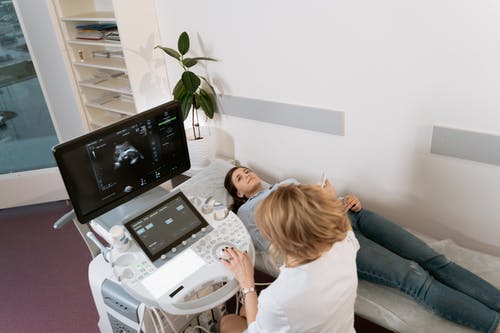
During a transabdominal ultrasound, you will lie on an examination table while the sonographer applies a special gel to your abdomen. The transducer will then be moved across your lower belly to capture images of your pelvic organs. You may feel slight pressure, but the procedure is painless.
Transvaginal Ultrasound:
For a transvaginal ultrasound, you will lie on your back with your knees bent. The sonographer will insert a lubricated transducer into the vagina to capture detailed images of the pelvic organs. This type of ultrasound provides clearer views of the ovaries and nearby structures, and while you may feel some pressure, the procedure is usually not painful.
After the ultrasound, the results will be analyzed by a radiologist or your doctor, who will explain the findings and discuss the next steps, if any.
Ultrasound Findings and Treatment Planning
If endometriomas or other features of endometriosis are identified on ultrasound, your healthcare provider will discuss treatment options. The treatment plan will depend on the severity of your symptoms, the location and size of the endometriomas, and whether you are trying to conceive.
Common Treatment Options Include:
- Medication: Hormonal therapies such as oral contraceptives, gonadotropin-releasing hormone (GnRH) agonists, or progestins are commonly prescribed to reduce the symptoms of endometriosis.
- Surgery: In some cases, surgery may be required to remove endometriomas, adhesions, or other growths. Laparoscopy is often used for both diagnosis and treatment, allowing surgeons to remove or burn away endometrial tissue.
- Fertility Treatment: For women with endometriosis-related infertility, fertility treatments like in vitro fertilization (IVF) may be recommended.
Follow-up and Monitoring with Ultrasound
Women with endometriosis may need regular follow-up ultrasounds to monitor the condition, especially if endometriomas are present. These ultrasounds help assess whether the cysts are growing and if there is a need for further intervention. Regular monitoring is also important if you are undergoing hormonal treatment or trying to conceive, as it allows your healthcare provider to adjust the treatment plan as needed.
Conclusion
Ultrasound plays an important role in diagnosing and managing endometriosis, particularly by detecting endometriomas and assessing pelvic anatomy. While it has limitations in visualizing all aspects of endometriosis, it is a non-invasive, widely accessible, and effective tool in the overall diagnostic process. Understanding how ultrasound fits into the broader management of endometriosis can help patients work closely with their healthcare providers to develop an effective treatment plan that addresses both symptoms and reproductive goals.