Vaginal probing technique
In the field of medical imaging, mastering vaginal transducer technology is paramount for healthcare professionals. The purpose of this article is to explore the optimization of ultrasound imaging, specifically for vaginal examinations, with a focus on improving patient care through better technology and interpretation.

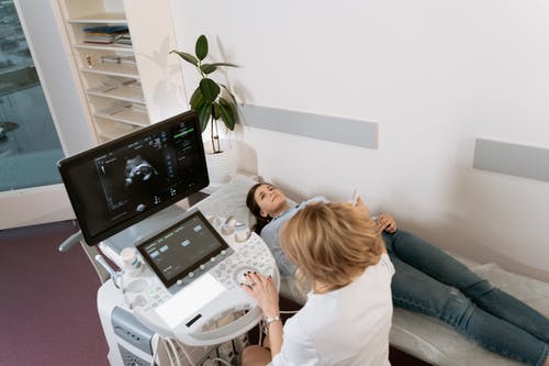
Vaginal ultrasound
Vaginal ultrasound, also known as transvaginal ultrasound, involves inserting a probe inside the vagina to take high-resolution images of the pelvic organs. Unlike an abdominal ultrasound, which is based on a probe placed outside the abdominal cavity, a vaginal ultrasound offers better image quality and better visualization of the internal structures of the pelvis.
Preparing for a vaginal ultrasound
Before having a vaginal ultrasound, it is important to ensure proper preparation. This includes adequately explaining the procedure to the patient, obtaining informed consent, and addressing concerns. In addition, healthcare providers should ensure that the ultrasound device is properly calibrated and the vaginal probe is properly sterilized and lubricated.
Optimizing Image Quality
Achieving the best possible image quality requires attention to detail and familiarity with ultrasound equipment. Healthcare providers must adjust settings such as frequency, gain, and depth to optimize viewing of the pelvic organs. Correct placement of the vaginal probe is also important to ensure proper contact with the vaginal walls to minimize artifacts and maximize image clarity.
Improve patient comfort
Patient comfort should be a priority for vaginal ultrasound examinations. Effective communication, empathy, and a supportive approach can help relieve anxiety and discomfort. Healthcare providers should take the time to address patient concerns and ensure the duration of the procedure.
Results
Accurate interpretation of ultrasound results is critical for patient management and treatment decisions. Health care providers must be able to identify normal anatomical structures and identify common conditions such as ovarian cysts, fibroids, and endometrial abnormalities. Clear documentation of findings is important for effective communication with other members of the healthcare team.
Case studies and examples
Real-life case studies can provide valuable information about the impact of proper technique and interpretation in vaginal ultrasound. By sharing examples of successful diagnoses and patient outcomes, healthcare providers can illustrate the importance of mastering vaginal sensor technology to improve patient care.
Continuous Improvement
Mastering vaginal sensor technology is an ongoing process that requires a commitment to continuous learning and improvement. Healthcare providers must actively seek educational opportunities, stay abreast of advances in ultrasound technology, and apply feedback and learning to practice.
Conclusion
To conclude, mastering the vaginal probing technique is vital for midwives to provide accurate and compassionate care. By honing this skill, midwives can ensure precise diagnostics and improve patient outcomes, enhancing the overall quality of maternal healthcare. Proficiency in this technique not only strengthens the trust between midwives and their patients but also empowers midwives to deliver the highest standard of care.